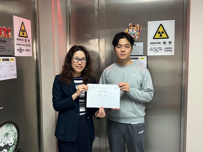
UM research team receives gold award for AI applications in medical image segmentation at East Asia Nuclear Medicine Association Congress
澳大團隊東亞核醫學聯合學術大會奪金獎


Mok Seng Peng, Greta (left) and Chen Gefei
莫昇萍(左)及陳戈飛

Award Certificate of East Asia Nuclear Medicine Association Congress
東亞核醫學聯合學術大會金獎證書
A research team from the Biomedical Imaging Laboratory (BIG), Faculty of Science and Technology (FST), University of Macau (UM) virtually attended the 3rd East Asia Nuclear Medicine Association Congress, hosted in Osaka, Japan in November. Competing with various institutes of nuclear medicine in Asia, including China Medical University, National Taiwan University Hospital, Taichung Veterans General Hospital, Tokyo Medical and Dental University, Kagawa University Kansai Medical University, and Osaka University, Chen Gefei, a PhD student from BIG lab, won the gold award in the meeting for his work on deep-learning-based segmentations of the liver and tumors from contrast-enhanced computed tomography (CECT) for Yttrium-90 radioembolization dosimetry. This advancement facilitates the treatment planning for hepatocellular carcinoma and liver metastases in Yttrium-90 radioembolization.
Yttrium-90 radioembolization is an effective treatment for inoperable liver tumors, and the treatment planning requires delineation of normal liver and tumors. Deep learning-based methods are feasible for tumor segmentation on single-phase CECT, but there are limitations in differentiating tumors and cysts. In Chen’s project entitled ‘Deep Learning-based Automated Segmentation of Liver and Tumors on CT Arterial Portography and Hepatic Arteriography for Yttrium-90 Radioembolization’, the group evaluated two deep learning-based methods on single-phase and dual-phase CECTs, respectively, for normal liver and tumor segmentation. The deep learning-based method using attention U-Net trained on dual-phase CECTs can achieve superior segmentation accuracy and successfully discriminate cysts and tumors. This method can highly facilitate the Y-90 radioembolization treatment planning procedure.
This project was led by Mok Seng Peng, Greta, a professor in the Department of Electrical and Computer Engineering, Institute of Collaborative Innovation, and the Ministry of Education Frontiers Science Center for Precision Oncology. It was a collaboration between UM and the Department of Nuclear Medicine, the Taipei Veterans General Hospital, and funded by Fundo para o Desenvolvimento das Ciências e da Tecnologia (file number: FDCT 099/2021/A).
澳門大學科技學院生物醫學成像實驗室(BIG)研究團隊近期於線上參加日本大阪舉行的“第三屆東亞核醫學聯合學術大會”。在與亞洲多個核醫學機構的競爭中,包括中國醫科大學和日本大阪大學等,BIG實驗室的博士生陳戈飛利用深度學習于增強CT圖像上進行肝臟和腫瘤分割,應用于釔-90放射栓塞劑量學研究,贏得了該大會的金獎。這一重要突破有望對肝細胞癌和肝轉移癌的病人在進行釔-90放射栓塞治療前提供更精准的治療計劃指導。
釔-90放射栓塞療法是治療難以手術切除的肝臟腫瘤的有效方法,治療計畫要求準確勾畫正常肝組織和腫瘤。基於深度學習的方法在單相增強CT圖像上進行腫瘤分割已經被證明是可行的,但在區分腫瘤和囊腫方面仍存在一定局限性。陳戈飛的研究,題為《基於CT動脈性門靜脈造影和肝動脈造影的深度學習自動肝臟和腫瘤分割應用於釔-90放射栓塞治療》,系統評估了兩種基於深度學習的方法在單相和雙相增強CT圖像上分別用於正常肝組織和腫瘤分割的效果。基於雙相增強CT圖像訓練的attention U-Net深度學習方法能夠實現較優越的分割準確度,並能成功區分囊腫和腫瘤。這一創新性方法將大大提高釔-90放射栓塞治療計畫的精准性與便利度,有利於病人個性化治療的實施。
該研究項目的指導教授為澳大電機及電腦工程系、協同創新研究院與精準腫瘤學前沿科學中心教授莫昇萍,合作單位為台北榮民總醫院核醫科,專案獲澳門科技發展基金資助(檔案編號:FDCT 099/2021/A)。