
News Express: UM achieves breakthrough in deep-brain angiography
新聞快訊:澳大在腦部深層血管造影技術取得新突破
低功率實現腦部深層血管造影術
Schematic illustration of low-power deep brain angiography and 3D reconstructions enabled by BTHG imaging
澳大在腦部深層血管造影技術取得新突破
澳門大學健康科學學院副教授劉子銘及研究助理教授徐昌活帶領的研究團隊聯合香港科技大學傑出客座教授唐本忠、研究教授林榮業的研究團隊,在腦部深層血管造影技術領域取得重大進展。研究團隊成功突破非線性光學成像技術,可在極低激光功率下獲取腦部深層(500微米)血管的高解析度影像,顯著降低診療過程中的光損傷風險,為早期篩查腦部微小動脈瘤等隱匿性腦血管病變提供了新方向。相關成果已發表於國際頂尖期刊《先進材料》(Advanced Materials)。
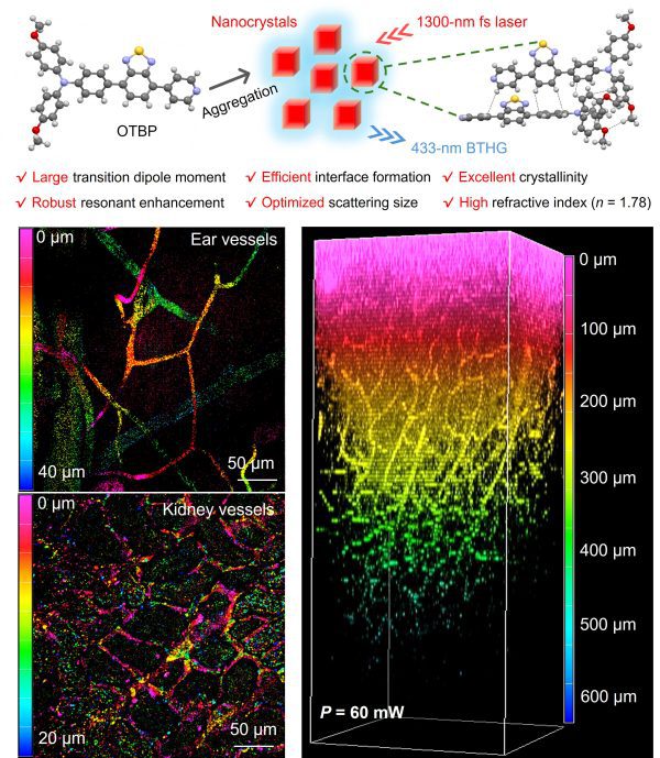
1960年,Theodore H. Maiman發明第一台紅寶石激光,開啓了激光技術的新紀元。科學家發現,當激光作用於石英晶體時,會產生一系列非線性光學(NLO)現象,其中三次諧波產生(THG)因其在介面處的信號增強特性備受關注。THG是一種四波和頻過程,如1300 nm的入射光可轉換為433 nm的THG。該技術憑藉高組織穿透深度和檢測靈敏度,被廣泛應用於生物組織的免標記成像。然而,傳統THG材料面臨效率低、背向信號收集困難、生物相容性差等瓶頸,難以應用於腦科學。若單純通過提高激光強度增強信號,又會引發組織自發螢光干擾和光損傷風險,導致體內成像研究長期停滯。如何平衡效率與安全性,成為領域內亟待解決的難題。
研究團隊設計了一系列有機小分子探針,通過分子層面的π-電子堆疊優化與介觀形貌調控,篩選出高效THG探針“OTBP”。實驗表明,OTBP可自組裝為高折射率(n=1.78)的穩定納米晶體,顯著提升背向THG信號收集效率。在僅60 mW低功率激光(脈衝能量0.75 nJ)激發下,該技術成功實現了小鼠腦部深層(500微米)血管的高對比成像,可清晰觀測直徑6.8微米的微小血管,信噪比高達143。與傳統CT及核磁共振血管造影相比,該技術兼具高分辨率與低損傷特性,有望彌補CT與核磁共振血管造影解析度低的缺點,與傳統血管造影術形成互補。該技術有望應用於腦卒中、動脈瘤等疾病的早期篩查,推動微創診療技術的發展。
該研究通訊作者為劉子銘、徐昌活、林榮業、唐本忠,澳大健康科學學院博士生杜立冬與香港科技大學博士沈翰辰為共同第一作者。澳大健康科學學院博士生周青青、葉栩昊及其它團隊成員也對研究作出重要貢獻。該研究由澳門特別行政區科學技術發展基金(檔案編號:0049/2023/ITP2、0003/2023/RIC、0013/2023/RIC)資助。全文章可瀏覽:https://advanced.onlinelibrary.wiley.com/doi/full/10.1002/adma.202414419。
欲瀏覽官網版可登入以下連結:
https://www.um.edu.mo/zh-hant/news-and-press-releases/campus-news/detail/61435/
UM achieves breakthrough in deep-brain angiography
A research team led by Associate Professor Liu Tzu-Ming and Research Assistant Professor Xu Changhuo in the Faculty of Health Sciences (FHS) at the University of Macau (UM), in collaboration with a team led by Distinguished Visiting Professor Tang Ben Zhong and Research Professor Lam Wing Yip at the Hong Kong University of Science and Technology (HKUST), has made significant progress in deep-brain angiography. The team has successfully optimised nonlinear optical (NLO) imaging technology to capture high-resolution images of the deep-brain vasculature (up to 500μm) using extremely low laser power. This significantly reduces the risk of photodamage during diagnostic procedures. This breakthrough offers a new approach to the early screening of subclinical cerebrovascular diseases, such as microaneurysms. The research has been published in the prestigious international journal Advanced Materials.
Theodore H. Maiman invented the first ruby laser in 1960, marking the dawn of laser technology. Scientists later discovered that when lasers interact with quartz crystals, a series of NLO phenomena occur. Among these, third-harmonic generation (THG) has drawn significant attention due to its signal enhancement properties at interfaces. THG is a four-wave mixing process that can convert 1,300nm incident light to a 433nm THG output signal. This technology is widely used for label-free imaging of biological tissue owing to its high tissue penetration depth and detection sensitivity. However, traditional THG materials have several limitations, including low efficiency, the difficulty of collecting backward signals, and poor biocompatibility. These limitations hinder their application in brain science. Furthermore, increasing the laser power to achieve satisfactory backward THG (BTHG) output can cause irreversible photodamage to tissue, which has long hindered progress in in vivo imaging research. Achieving a balance between efficiency and safety remains a critical challenge in the field.
To address these limitations, the research team designed a series of organic small-molecule probes. By optimising the molecular packing of π-electron and controlling the mesoscale morphology, the team identified an efficient THG probe named ‘OTBP’. Results demonstrated that OTBP self-assembled into stable nanocrystals with a high refractive index (n=1.78), significantly enhancing BTHG signal output. Under low-power laser excitation (60mW, pulse energy 0.75nJ), the technology achieved high-contrast imaging of deep-brain vasculature (500μm) in mice. This enabled the visualisation of microvessels as small as 6.8μm in diameter, with a signal-to-noise ratio of 143. Compared to traditional CT and MRI angiography, this new technology offers high resolution and minimal invasiveness. It can address the issue of low resolution in conventional methods and complement existing angiography techniques. The technology also shows promise in the early detection of conditions such as stroke and aneurysms, advancing the development of minimally invasive diagnostics.
The corresponding authors of the study are Liu Tzu-Ming Liu, Xu Changhuo, Lam Wing Yip, and Tang Ben Zhong. The co-first authors are Du Lidong, a doctoral student from UM FHS, and Shen Hanchen, a doctoral graduate from HKUST. UM doctoral students Zhou Qingqing and Ye Xuhao, along with other team members also made significant contributions to the study. The research was funded by the Science and Technology Development Fund of the Macao SAR (File No.: 0049/2023/ITP2, 0003/2023/RIC, 0013/2023/RIC). The full version of the research article is available at: https://advanced.onlinelibrary.wiley.com/doi/full/10.1002/adma.202414419.
To read the news on UM’s official website, please visit the following link:
https://www.um.edu.mo/news-and-press-releases/campus-news/detail/61435/