
News Express: UM research team develops innovative wearable optical brain functional imaging device
新聞快訊:澳大團隊成功開發創新可穿戴光腦功能成像儀
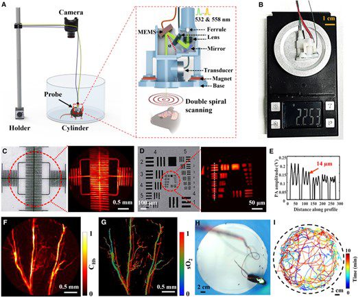
系統組態與性能表徵
System configuration and performance characterisation
澳大團隊成功開發創新可穿戴光腦功能成像儀
澳門大學認知與腦科學研究中心主任、健康科學學院教授袁振帶領的研究團隊成功開發創新可穿戴光聲血流代謝顯微鏡(PHM),不僅適用於卒中研究,還可拓展至阿爾茨海默病、帕金森病等神經退行性疾病機制探索,對精準醫學和神經疾病研究具有重要意義。相關研究成果已發表於國際頂尖期刊《Cell Reports Physical Science》。
腦卒中是危害極大的腦血管疾病,動物卒中模型是研究卒中機制和治療方法的重要手段。然而,傳統模型需麻醉或固定動物,難以呈現真實生理過程。PHM突破這一限制,可在自由運動的小鼠中精準誘導特定區域血管栓塞並即時監測血氧飽和度與血紅蛋白濃度,捕捉血栓形成及神經血管代謝耦合的動態過程。
研究結果顯示,單血管栓塞並非線性阻斷,而呈現“堵塞—再通—再堵塞”的動態過程,揭示血管具備一定的血栓清除能力。伴隨血紅蛋白含量和血氧飽和度下降,小鼠活動逐漸減少,在重度栓塞模型中,研究團隊觀察到血管結構和功能連接發生重塑,血氧飽和度出現階段性提升,但嚴重栓塞仍導致高死亡率。研究團隊還建立輕度栓塞模型,進行28天縱向監測,呈現血管再通、運動能力恢復及腦功能連接重組的全過程。
該技術為卒中模型構建及腦疾病研究提供全新平台,實現自由活動小鼠腦血管栓塞過程的即時、高解析度成像,突破傳統成像技術限制。PHM系統採用雙波長激發與光聲檢測技術,重量不足2.3克,具備8 mm²的成像視野和330毫秒的時間解析度,適用於卒中研究,以及探索阿爾茨海默病、帕金森病等神經退行性疾病的機制。
該研究通訊作者為袁振,第一作者為澳大健康科學學院博士畢業生梁笑,南方科技大學教授奚磊亦對研究作出貢獻。該研究獲深圳市醫學研究基金(檔案編號:B2402046)、澳門大學(檔案編號:MYRG2022-00054-FHS,MYRG-GRG2023-00038-FHS-UMDF,MYRG-GRG2024-00259-FHS)、澳門特別行政區科學技術發展基金(檔案編號: 0014/2024/RIB1)、國家自然科學基金項目(檔案編號:62435008)、深圳市科技計劃項目(檔案編號:RCJC20231211090039066,JCYJ20230807093105010)、南方科技大學創業基金(檔案編號:PDJH2021C008)資助。全文可瀏覽:https://www.cell.com/cell-reports-physical-science/fulltext/S2666-3864(25)00555-7。
欲瀏覽官網版可登入以下連結:
https://www.um.edu.mo/zh-hant/news-and-press-releases/campus-news/detail/63092/
UM research team develops innovative wearable optical brain functional imaging device
A research team led by Yuan Zhen, head of the Centre for Cognitive and Brain Sciences and professor in the Faculty of Health Sciences (FHS) at the University of Macau (UM), has developed an innovative wearable photoacoustic hemometabolic microscope (PHM). In addition to stroke research, the PHM system can be extended to studies of neurodegenerative disorders such as Alzheimer’s and Parkinson’s disease, offering significant implications for precision medicine and neuroscience. The findings have been published in the leading international journal Cell Reports Physical Science.
Stroke is a major cerebrovascular disease with severe consequences. Animal stroke models are essential for studying underlying mechanisms and therapeutic strategies. However, conventional approaches require anaesthesia or head fixation, which limits the ability to capture authentic physiological processes. The PHM overcomes these limitations by enabling precise induction of vascular occlusion in freely moving mice while simultaneously monitoring blood oxygen saturation and haemoglobin concentration in real time, thereby capturing thrombus formation and neurovascular-metabolic coupling dynamics.
The study reveals that single-vessel occlusion does not occur linearly but follows a dynamic ‘occlusion–recanalisation–reocclusion’ pattern, indicating that vessels possess a certain capacity for thrombus clearance. As haemoglobin content and oxygen saturation decline, mice exhibit progressively reduced locomotor activity. The team observed that, in severe occlusion models, vascular structural remodelling and functional connectivity changes, along with transient improvements in oxygen saturation, although mortality remained high. A mild occlusion model was also established for 28-day longitudinal monitoring, documenting vascular recanalisation, motor recovery, and functional network reorganisation.
This technology provides a novel platform for stroke modelling and brain disease research, enabling real-time, high-resolution imaging of vascular occlusion in freely moving animals and overcoming the limitations of traditional imaging techniques. The PHM system employs dual-wavelength excitation and photoacoustic detection, weighs less than 2.3 g, and offers a field of view exceeding 8 mm² with a temporal resolution of 330 ms. Beyond stroke research, the PHM system can be extended to studies of neurodegenerative disorders such as Alzheimer’s and Parkinson’s disease.
Yuan Zhen is the corresponding author of the study, with Liang Xiao, a doctoral graduate of UM FHS, as the first author. Xi Lei, professor at the Southern University of Science and Technology, also contributed to the study. The research was supported by the Shenzhen Medical Research Fund (File No: B2402046); UM (File Nos: MYRG2022-00054-FHS, MYRG-GRG2023-00038-FHS-UMDF, MYRG-GRG2024-00259-FHS); the Macao Science and Technology Development Fund (File No: FDCT 0014/2024/RIB1); the National Natural Science Foundation of China (File No: 62435008); the Shenzhen Science and Technology Program (File Nos: RCJC20231211090039066, JCYJ20230807093105010), and a start-up grant from the Southern University of Science and Technology (File No: PDJH2021C008). The full version of the research article is available at: https://www.cell.com/cell-reports-physical-science/fulltext/S2666-3864(25)00555-7.
To read the news on UM’s official website, please visit the following link:
https://www.um.edu.mo/news-and-press-releases/campus-news/detail/63092/