News Express: UM Stanley Ho East Asia College, Xinya College of Tsinghua University become sister colleges
jasonleong2025-05-13T17:52:33+08:00
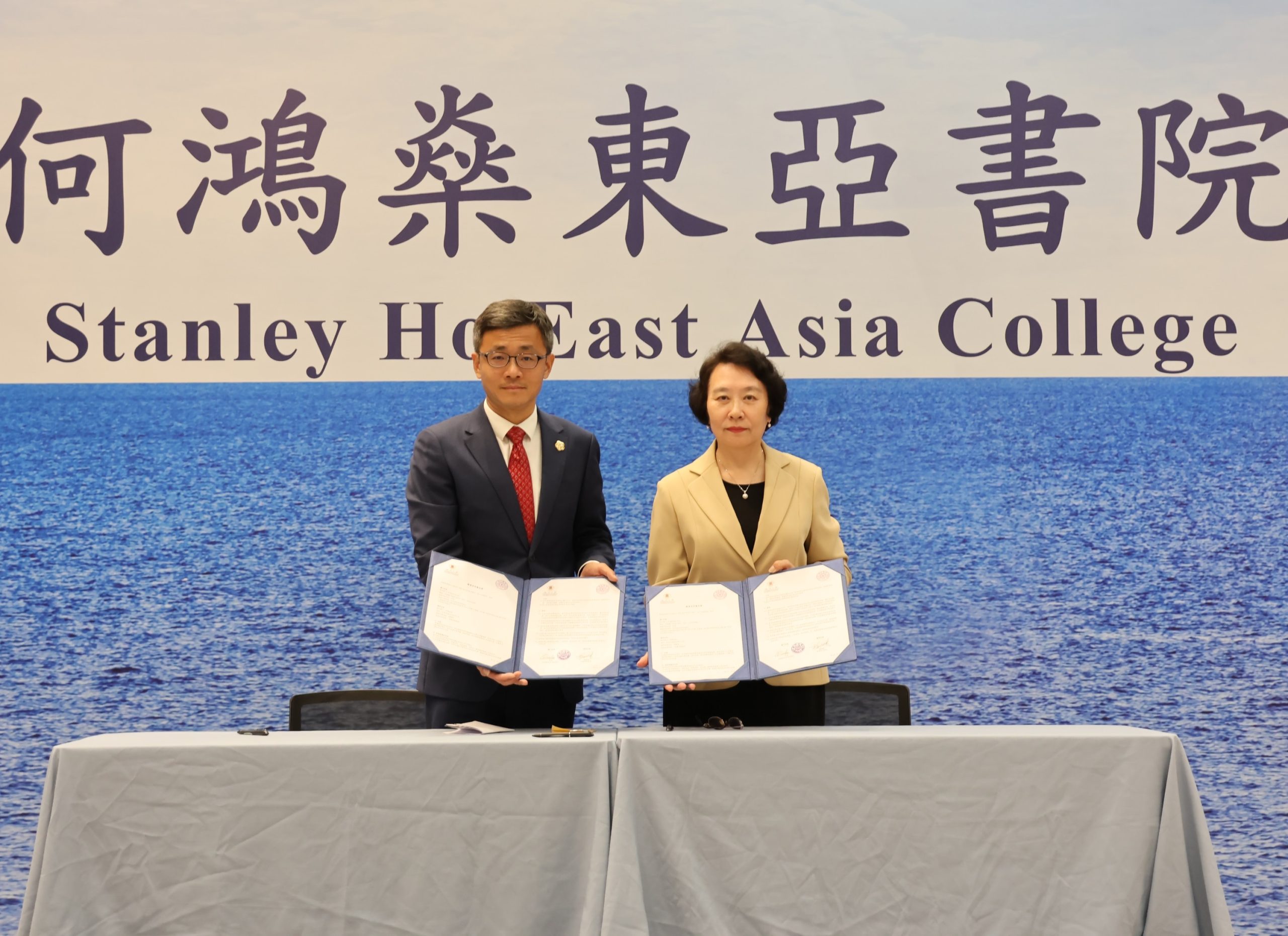
雙方簽署教育合作備忘錄
The signing of the memorandum of understanding on educational cooperation
澳大何鴻燊東亞書院與清華新雅書院締結姊妹書院
澳門大學何鴻燊東亞書院與清華大學新雅書院在2025年4月29日締結為姊妹書院,將從多方面開展兩院的交流和合作。
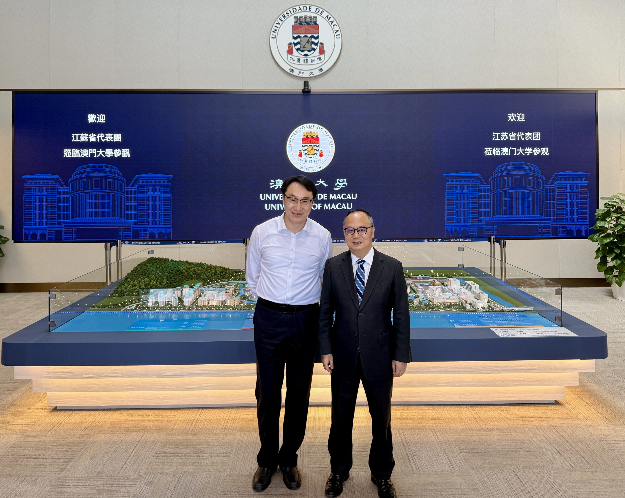
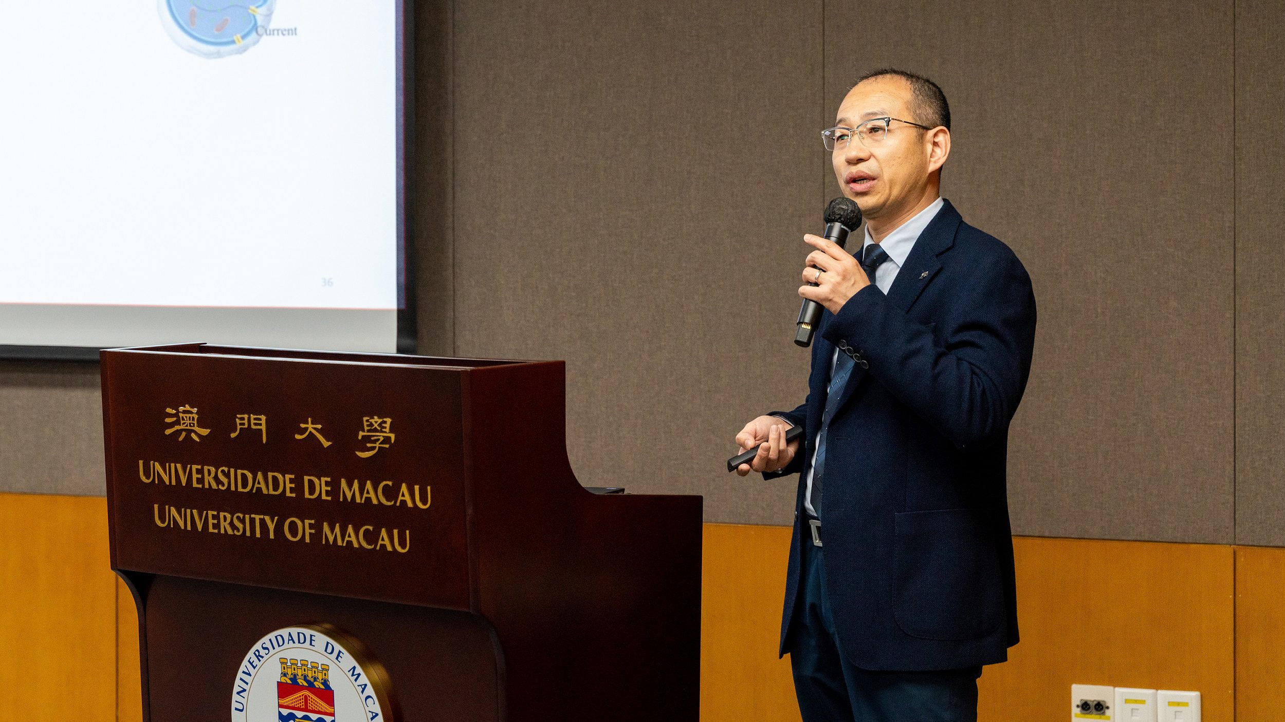
清華新雅書院師生一行到訪澳大。期間,在澳大副校長莫啓明見證下,澳大何鴻燊東亞書院院長羅茜與清華新雅書院院長梅賜琪簽署了教育合作備忘錄,正式締結為姊妹書院。雙方將推動師生互訪,加強兩院交流和合作,攜手探索書院制教育模式的創新發展。
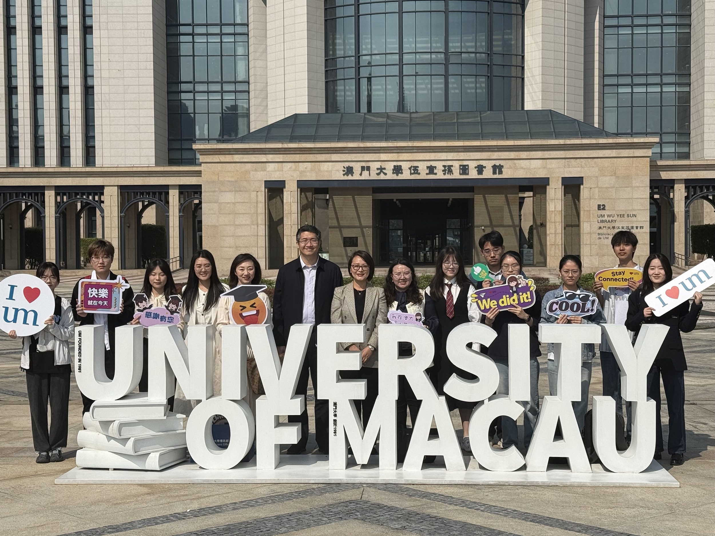
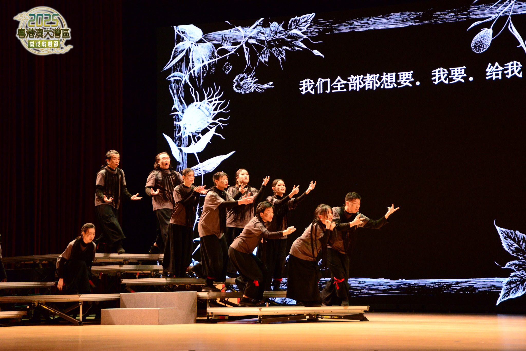
清華師生一行還參觀了澳大校園及澳門歷史城區,以及參加了澳大何鴻燊東亞書院舉行的2024—2025年度畢業高桌晚宴,約250名師生共聚一堂,一同分享畢業生的喜悅。梅賜琪表示,是次交流了解到澳大何鴻燊東亞書院在教育及文化建設方面的長遠規劃和精細執行,是一次寶貴的書院教育探索經驗。
澳大何鴻燊東亞書院是澳大推行“四位一體”教育模式而建立的首批住宿式書院;清華新雅書院則是清華大學為探索世界一流本科教育而設立的第一所住宿制書院。
欲瀏覽官網版可登入以下連結:
https://www.um.edu.mo/zh-hant/news-and-press-releases/press-release/detail/61128/
UM Stanley Ho East Asia College, Xinya College of Tsinghua University become sister colleges
Stanley Ho East Asia College (SHEAC) of the University of Macau (UM) and Xinya College of Tsinghua University have established a sister-college relationship, marking the beginning of academic and cultural exchanges between the two colleges.
The partnership was formalised through the signing of a memorandum of understanding (MOU) on educational cooperation during a visit to UM by a delegation from Xinya College. The MOU was signed by Luo Qian, college master of SHEAC, and Mei Ciqi, dean of Xinya College, under the witness of Mok Kai Meng, vice rector of UM. Under the MOU, the two parties will foster student and faculty exchanges, strengthen cooperation, and promote innovative developments in residential college education models.
During their time in Macao, the Tsinghua delegation toured the UM campus and the Historic Centre of Macao. They also attended SHEAC’s valedictory dinner for the 2024/2025 academic year.
To read the news on UM’s official website, please visit the following link:
https://www.um.edu.mo/news-and-press-releases/press-release/detail/61128/